Phages, which live in the digestive tract, could treat bacterial infections if issues impacting effectiveness are solved. Research at UT Health San Antonio and UTSA is doing just that.
Could beneficial viruses that live in the gut replace or supplement antibiotics to treat bacterial infections such as meningitis, botulism and E. coli? Researchers from The University of Texas Health Science Center at San Antonio (also called UT Health San Antonio) and The University of Texas at San Antonio are studying this with a grant from the San Antonio Medical Foundation.

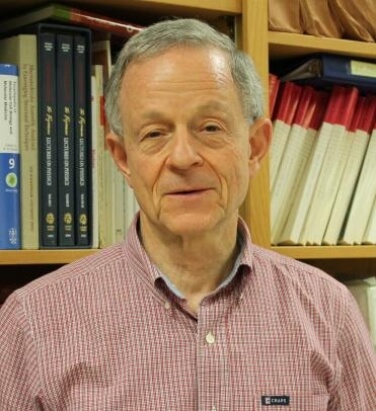
The viruses are called phages and have been proposed as therapy for bacterial infections since the 1920s. “In fact, phages were the original antibiotics before chemical antibiotics, such as penicillin, were developed,” said Philip Serwer, PhD, professor of biochemistry and structural biology at UT Health San Antonio. “Their use as antibiotics went down with the rise of chemical antibiotics.”
Resistant infections: Tip of the iceberg
Over time, though, bacteria develop resistance against powerful chemical antibiotics. The U.S. Centers for Disease Control and Prevention (CDC) estimates 2.8 million antimicrobial-resistant infections occur each year, and more than 35,000 people die of these infections annually. “Articles suggest that the number of deaths is actually much higher than that,” Serwer said. “Multiple-drug-resistant infections may be much more common than people realize.”
Enter phages. For each bacterium, there are corresponding phages that will kill it. Our intestines are replete with these cohabitating viruses, with many thought to have a positive effect. “No phage has ever been found to be toxic to humans,” Serwer said. And there are instances in which phages saved people who were dying from bacterial infections.
Some phages are quickly erased
Given these positives, why was phage therapy phased out in favor of chemical antibiotics? To put it simply, a key discovery made at UT Health San Antonio is that some, but not all, phages are removed by white blood cells before they can be effective. “A key objective of our research is rapidly finding the phages that are removed slowly and to keep all phages working for as long as possible,” Serwer said. “The key problem is getting phage therapy to work more consistently.”
In previous research funded by the Robert J. Kleberg, Jr. and Helen C. Kleberg Foundation, Serwer and colleagues solved problems with quickly and inexpensively isolating, characterizing and purifying phages in needed amounts. The issue of phages entering the bloodstream and being taken out by white blood cells is the major remaining hurdle.
Mouse studies
Experiments with collaborator Cara Gonzales, DDS, PhD, of the UT Health San Antonio School of Dentistry tested four different phages and looked at how long they stayed in the blood of the same mouse.
“We found that some phages persisted while others did not,” Serwer said, noting that the identification of phages that have this trait of high persistence is critical for potential therapeutics.
Electrical charge
What underlies this trait? How do other large objects, such as red blood cells, persist in the blood? In the case of red blood cells, they have a very high negative electrical charge on them, Serwer said.
“This leads us to the hypothesis that maybe the phages that have the highest persistence also have the highest negative electrical charge on their surfaces, making them excellent candidates to treat drug-resistant bacteria,” Serwer said. “This is the basis of our San Antonio Medical Foundation-supported research.”
The team is sifting 400 phages, many preserved from the Kleberg Foundation-funded research, to identify the best therapeutic candidates.
Practical issue solved
Serwer developed a collaboration with former UT Health San Antonio graduate student James P. Chambers, PhD, to develop chemical procedures that kill infectious bacteria without harming beneficial phages. Chambers, now a professor in the Department of Molecular Microbiology and Immunology at The University of Texas at San Antonio, succeeded in doing this, which solves another practical issue in characterizing phages grown in disease-causing bacteria for the prospective advent of high-performance phage therapy.
Acknowledgments
In the project, funded by $100,000 from the San Antonio Medical Foundation, Philip Serwer, PhD, Department of Biochemistry and Structural Biology at UT Health San Antonio, joined forces with James P. Chambers, PhD, Department of Molecular Microbiology and Immunology at The University of Texas at San Antonio, and Cara Gonzales, DDS, PhD, Department of Comprehensive Dentistry, UT Health San Antonio. Key participants are Elena T. Wright of the Serwer laboratory and Jorge de la Chapa of the Gonzales laboratory, along with students Samantha Roberts, Miranda Aldis and Meagan Weaver-Rosen from the Department of Microbiology, Immunology and Molecular Genetics at UT Health San Antonio. The structure of bacteria killed by the Chambers procedure was determined by electron microscopy assisted by Barbara Hunter in the Department of Pathology, UT Health San Antonio. Serwer and Chambers recently joined with a business colleague, William Tolhurst, to form a startup company, called Phage Refinery LLC, to optimally implement the phage research findings.


