SAN ANTONIO (Dec. 7, 2011) — A cardiologist-inventor from UT Medicine San Antonio who is devising innovative uses for a light-based technology with potential to predict, or even prevent, heart attacks will become an investigator for the Houston-based Clayton Foundation for Research, continuing his work with the foundation’s support.
UT Medicine San Antonio
Marc D. Feldman, M.D., and his longstanding collaborator, biomedical engineer Thomas E. Milner, Ph.D., of the Cockrell School of Engineering at The University of Texas at Austin, are at the forefront of bringing the technique into the practice of cardiology.
Becoming Clayton investigators will allow them to refine the technology so it can identify one more hallmark of a plaque vulnerable to rupturing within blood vessels. Ruptures of these fatty deposits are the most common cause of heart attacks and strokes, leading causes of death worldwide.
The UT Medicine San Antonio project is one of six new medical research programs being established by the Clayton Foundation in Texas in 2012. Each program initially will receive $300,000 per year not only to support investigators like Drs. Feldman and Milner, but also to add postdoctoral fellows and cover equipment and supplies. The Clayton Foundation and its supporting organization, the Foundation for Research, have supported medical research within The University of Texas System since 1933.
“The Clayton Foundation allows its investigators wider latitude in their research than traditional grant-funding agencies, such as the National Institutes of Health,” said Dr. Feldman, who sees patients through UT Medicine San Antonio, the practice group of the School of Medicine at The University of Texas Health Science Center at San Antonio. “As a result, rather than focusing on research that will lead to the next funded grant application, investigators can focus on what is most exciting and most likely to impact patient care.”
Drs. Feldman and Milner collaborate with other investigators, including chemical engineer Keith P. Johnston, Ph.D., from The University of Texas at Austin, and biochemist Reto Asmis, Ph.D., at the UT Health Science Center. Together, these four investigators have had great success in developing cardiovascular applications for the light-based technique, called Optical Coherence Tomography (OCT).
OCT harnesses light waves instead of the sound waves, magnetic fields and radiation now used to image cardiovascular disease. OCT has far greater sensitivity, specificity and resolution than any of those techniques, with none of the harmful effects of radiation.
The researchers’ previous work in OCT resulted in seven U.S. patents and a spin-off company in 2005. The intellectual property and all seven patents are managed through the office now called South Texas Technology Management (STTM), which assists in commercializing technologies developed at five Texas universities, including the UT Health Science Center.
The spin-off company, CardioSpectra, was financed in part by the Texas Emerging Technology Fund and remains the fund’s most profitable investment to date. When San Diego-based Volcano Corp. purchased CardioSpectra in 2007 for $25 million cash, plus the opportunity to earn $38 million in milestone payments, the state received cash and Volcano stock. The state will not fully realize its profit until the stock is sold, but at its 2011 peak, the fair market value of the state’s investment reflected a 260 percent gain.
Volcano received CE Mark in Europe on that first-generation system in December 2009 and is developing a second-generation system for widespread commercialization.
A relatively new technology, OCT emerged from laboratories at the Massachusetts Institute of Technology in the 1990s. Its earliest medical imaging applications were in ophthalmology; Drs. Feldman and Milner have been pioneers in bringing it to cardiology.
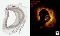
OCT involves directing near-infrared light at tissue. Most light scatters, but small amounts reflect back and can be used to form an image. The scattered light is filtered out.
The OCT technique created by the University of Texas System collaborators currently can detect two of three classic features of a plaque that is vulnerable to rupture: thinning of the tissue layer – or “fibrous cap” – that covers the plaque’s core, and an especially large collection of lipids beneath that cap.
The Clayton Foundation’s investment will allow researchers to tackle the third and final key characteristic of vulnerable plaques: an increase in the number of “macrophages,” or scavenger cells that digest pathogens and cellular debris. Macrophages have been shown to weaken the fibrous cap.
The OCT technique developed by the UT System team involves guiding a catheter through blood vessels – similar to intravascular ultrasound, which is already used in cardiac catheterization laboratories. While ultrasound can image plaques that are not visible through other methods, images are not detailed enough to identify those features of a plaque that predict heart attacks.
Because light vibrations are so much finer than sound, OCT produces images of far higher resolution than ultrasound, capturing details previously seen only at autopsy. OCT is in its infancy and, as the technology matures, will be an important tool for studying the biology of atherosclerosis, or hardening of the arteries, in patients for the first time.
“OCT is one of the most promising and exciting medical imaging technologies available today,” Dr. Milner said. “The detail of information OCT can provide is simply astonishing and will continue to impact not only cardiology but other medical disciplines, such as ophthalmology.”
Although the ability to predict heart attacks is still a ways off, a more imminent use for OCT might be determining whether the cells lining the walls of blood vessels have sufficiently repaired themselves after a drug-eluting stent is inserted. When this does not happen, patients are thought to be at risk for clots that obstruct the flow of blood and ultimately cause heart attacks. OCT can see this; ultrasound cannot.
UT Medicine San Antonio is the clinical practice of the School of Medicine at the UT Health Science Center at San Antonio. With more than 700 doctors – all faculty from the School of Medicine – UT Medicine is the largest medical practice in Central and South Texas, with expertise in more than 60 different branches of medicine. Primary care doctors and specialists see patients in private practice at UT Medicine’s clinical home, the Medical Arts & Research Center (MARC), located in the South Texas Medical Center at 8300 Floyd Curl Drive, San Antonio 78229. Most major health plans are accepted, and there are clinics and physicians at several local and regional hospitals, including CHRISTUS Santa Rosa, University Hospital and Baptist Medical Center. Call (210) 450-9000 to schedule an appointment, or visit the Web site at www.UTMedicine.org for a complete listing of clinics and phone numbers.
South Texas Technology Management (STTM) is the UT technology transfer office serving UT Health Science Center San Antonio, UT San Antonio and other institutions in the South Texas region. STTM provides leadership in promoting innovation and technology transfer through proactive management of IP, technology development and commercialization to support the missions of member institutions, advance regional economic development and benefit the public. Please refer to www.utsttm.org to learn more about STTM. To find other innovative UT technologies, visit http://www.utsystem.edu/sttm/technologies.shtml.