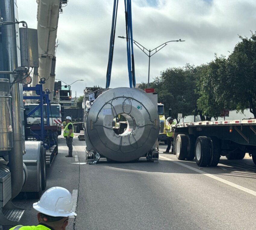
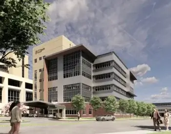
A leap forward in brain imaging is coming to San Antonio. UT Health San Antonio’s Center for Brain Health opening in December 2025 will be the first in Texas to house a Siemens MAGNETOM 7-Tesla Terra.X magnetic resonance imaging (MRI) system — one of the most advanced MRI technologies available. This scanner produces ultra-high-resolution brain images, offering a new tool for diagnosing and studying conditions like dementia, Alzheimer’s, Parkinson’s and other brain diseases.
The 7T Terra.X is an upgrade over conventional MRI scanners, which generally operate at 1.5 or 3 Tesla. MRI power is measured in Tesla units, with 7 Tesla representing a magnetic field strength more than 140,000 times that of Earth.

“This will improve detection of small metastases, multiple sclerosis lesions and more,” said Ray Lee, PhD, radiologist with UT Health San Antonio and associate professor of radiology in the Joe R. and Teresa Lozano Long School of Medicine at The University of Texas at San Antonio. “It does not replace the other MRIs in (our) Imaging Center and the Medical Arts and Research Center but rather expands what is possible.”
Compared to standard MRI, the 7T offers dramatically improved resolution, especially in deep or complex regions such as the brain stem and temporal lobes. It can distinguish differences that may not be visible with lower-powered scans. For example, the 7T can more accurately distinguish between a benign blood vessel and an aneurysm.
“This MRI lets us see incredible detail, even without contrast, including deep structures like the brain stem where many diseases begin, and veins where amyloid is cleared,” said Sudha Seshadri, MD, DM, neurology professor at UT San Antonio and founding director of the Glenn Biggs Institute for Alzheimer’s and Neurodegenerative Diseases at UT Health San Antonio, which will move to its new home at the UT Health San Antonio Center for Brain Health in December.
The scanner incorporates dynamic parallel transmit architecture, which eliminates image distortion caused by magnetic field inhomogeneity — a common challenge in ultra-high-field MRI.
“With eight transmitters instead of two, we can dynamically adjust the magnetic field to create a more uniform image, allowing us to clearly see regions that used to be obscured,” Lee said. “This has become the new standard for 7T imaging.”
The system can also switch between research and Food and Drug Administration-approved clinical modes in minutes.
“We’re right at the transition point where 7T MRI is becoming a clinical tool,” Lee said. “Currently we are mostly research-focused, but we will eventually be able to scan patients with much greater accuracy.”
This flexibility is already enabling work on surgical planning for deep brain stimulation, substance use disorder research and advanced diagnostics.
“This technology expands what is possible,” Lee said. “It will enhance research and discovery, right here in our community.”