UT Health San Antonio invests in state-of-the-art cryo-electron microscopy, adding a powerful new tool to solve disease riddles at the molecular level.
Contact: Will Sansom, 210-567-2579, sansom@uthscsa.edu
SAN ANTONIO (Aug. 18, 2022) — When physicians observe or hear of symptoms in their patients, they order CT scans, MRIs or other types of imaging analysis to understand the causes.
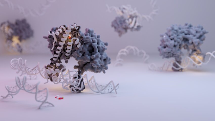
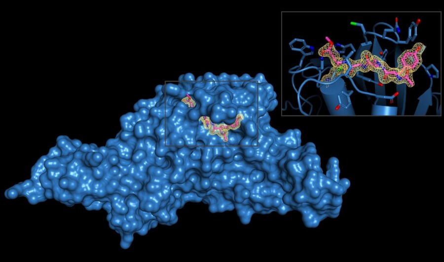
Clinical imaging, however, does not illuminate the vast machinations of life that occur deeper within, at the Lilliputian level of individual proteins and other molecules. Chain reactions in this tiny domain determine whether good health continues, or diseases begin. To explore how the reactions foster disorder, scientists acquire images with state-of-the-art instruments capable of ultra-high resolution. They employ statistical models to work out molecular structures. The overall analysis yields sites of disease susceptibility that can be targets for drug therapies.
UT Health San Antonio is investing $5 million over the next three years in such a technology, called cryo-electron microscopy, or cryo-EM for short. It is the hottest realm in structural biology today.
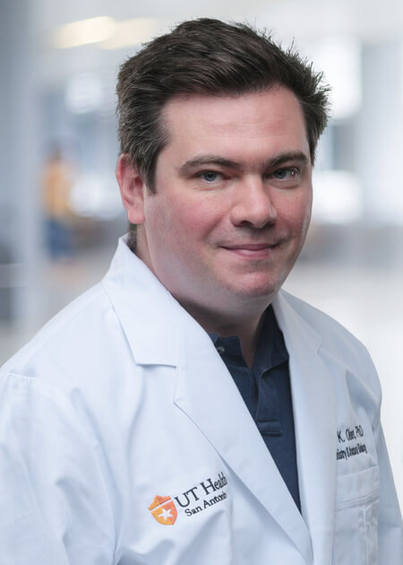
“We are essentially molecular photographers,” said Shaun Olsen, PhD, associate professor of biochemistry and structural biology and director of structural biology cores at UT Health San Antonio. “Some people take pictures of buildings. We take pictures of proteins and want to see what they look like in three dimensions.”
Cryo-EM visualizes proteins that are extremely difficult to image using other techniques, said Elizabeth Wasmuth, PhD, assistant professor of biochemistry and structural biology. She joined UT Health San Antonio this year after participating in cryo-EM studies of prostate cancer conducted at multiple institutions in New York.
Cryo-EM is complementary to existing structural biology technologies at UT Health San Antonio. X-ray crystallography, for example, exposes a protein crystal to X-rays, diffracting the X-ray beam in directions according to the protein’s structure. Nuclear magnetic resonance (NMR) spectroscopy, meanwhile, demonstrates behavior of an atom nucleus when it is placed in a powerful magnetic field. Experts can infer structure from the behavior they observe.
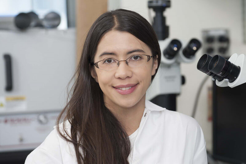
“We look at molecules in levels of detail that are unparalleled,” Dr. Wasmuth said. “So, basically, it is like being able to see a dime on the surface of the moon. This is the level of resolution that the techniques and tools of structural biology allow us to see about molecules inside cells, inside our bodies. Cryo-EM adds a powerful new dimension to our other methods.”
Some protein targets are too small to be visualized by existing techniques or have flexible, wiggly regions that impede the crystal formation, Dr. Wasmuth said. Cryo-EM flash-freezes proteins on thin layers of ice within milliseconds and barrages them with electron beams, generating biologically useful information.
“Having a cryo-EM system will allow us to observe drug targets that couldn’t be visualized by the other methods,” Dr. Wasmuth said. “The second week the cryoEM facility became functional, we were able to solve the structure of a complex of proteins involved in DNA damage repair at impressively high resolution. I am confident that this tool is going to transform structural biology research here like we haven’t seen before.”
Placing priority on the science that will cure disease
“The UT Health Science Center at San Antonio is a premier biomedical research enterprise because of its commitment to these types of investments,” said Jennifer Sharpe Potter, PhD, the institution’s vice president of research. “This is the latest in a long tradition of conscious, intentional decisions to maintain cutting-edge instruments required to answer questions that will translate science into practice.”
Patrick Sung, DPhil, who in 2020 was interim chairman of biochemistry and structural biology and now directs the Greehey Children’s Cancer Research Institute at UT Health San Antonio, collaborated with Dr. Olsen to plan and procure the cryo-EM system. Dr. Sung received the enthusiastic support of Dr. Potter and Robert Hromas, MD, vice president for medical affairs and dean of the Joe R. and Teresa Lozano Long School of Medicine. “We look forward to the results that will come,” Dr. Hromas said. “It is a significant investment not just in an instrument, but in scientists who will want to join us in San Antonio because of it.”
Separate multimillion-dollar awards made by the Cancer Prevention and Research Institute of Texas (CPRIT) aided the scientists’ recruitments. Dr. Olsen joined UT Health San Antonio from the Medical University of South Carolina, and Dr. Wasmuth was recruited from the Memorial Sloan Kettering Cancer Center and the Rockefeller University in New York City.
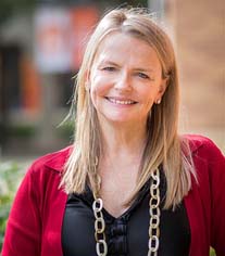
Funding from The University of Texas System Science and Technology Acquisition and Retention (STARs) program also aided the researchers with renovating their laboratories and purchasing instrumentation. STARs support has long been vital to UT Health San Antonio. “We’ve had an amazing year (Fiscal 2022) with STARs,” Dr. Potter said. “We received the highest level of recruitment awards from STARs that this institution has ever had in one year. It is more than $5 million, and we thank the UT System Board of Regents and STARs administrators for this.”
Planning and installation: The agony and the ecstasy
Dr. Olsen developed a business model for the cryo-EM facility in the summer of 2020. “We ordered it in December 2020, it took the manufacturer a year to build it, they shipped it around Thanksgiving in 2021 and it arrived here in early 2022,” he said. “In March 2022 renovations to a campus space were completed, and a team reassembled it and installed it.”
Choosing the space was a painstaking task.
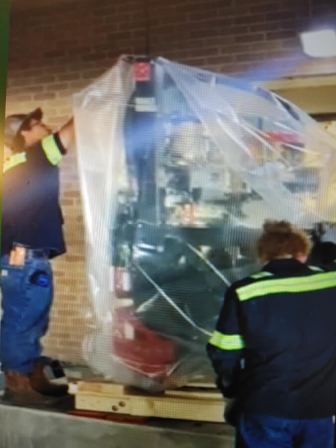
“A cryo-EM system is super-super sensitive, and we had to go through a process with the facilities and maintenance people to make sure there was no electromagnetic interference or vibrational interference,” Dr. Olsen said.
Dr. Potter had identified a place for the installation and “her intuition was on the mark, because the vendor said, ‘We’ve never seen scores this good,’” Dr. Olsen said.
During the site survey, Miguel Rivera, electrical engineer in the Department of Facilities Management, drove a truck multiple times through a parking garage that houses a door leading to the installation space. Would the vibrations be an issue? “It was COVID, so the traffic wasn’t what you normally see,” Dr. Wasmuth said. “Miguel had to drive by many times.”
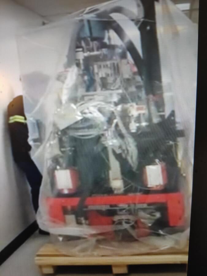
Once the cryo-EM system arrived, getting it into the building required ingenuity. The company shipped it in two pieces, but even so, the giant pieces cleared the door space and hallway by mere inches. “It was like doing a jigsaw puzzle to get it into the perfect space,” Dr. Wasmuth said.
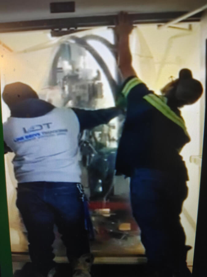
Raymundo Rivera, MBA, MS, PE, CxA, executive director of engineering and construction management and chief electrical engineer, Facilities Management, was also invaluable, the scientists said.
The process of acquiring such an instrument is arduous as a rule, and was especially challenging in 2020, the period of the COVID-19 pandemic before vaccinations became available.
“Getting people on campus in the beginning with the restrictions, knowing who could come to campus and when, slowed things down a bit,” Dr. Olsen said.
After installation, scientists are charged with making sure that an instrument is working as advertised. That process is ongoing. “It’s like if you get a car and it says it will go from zero to 60 in four seconds,” Dr. Olsen said. “Right now, we are seeing if the cryo-EM can go from zero to 60 in four seconds. This is not atypical for a very complex instrument. There is just troubleshooting.”
“It’s more common that you run into some kind of problem than you don’t,” Dr. Wasmuth said.
Because the microscope is so sensitive to vibrations and electromagnetic interference from air conditioners, centrifuges and other sources, the scientists needed a particular type of ceiling tile in the room. Delivery of the tiles was delayed by a few weeks during the pandemic because of lack of supply.
Through it all, the cryo-EM’s new digs, which are under the Holly Auditorium on campus, proved to be ideal. And they were selected with the future in mind.
“The space Dr. Potter chose for us is big enough so that we can expand the facility and eventually acquire another microscope,” Dr. Wasmuth said. “That shows the institutional commitment to the growth of the structural biology center.”
Big (very big) data
One dataset from two days on the cryo-EM system will be about 5 terabytes, Dr. Wasmuth said. A terabyte is 1 trillion bytes of information. This is clearly not something grandma’s computer can handle. It requires a graphic processing unit (GPU) to render graphics at high speed.
“We need very, very powerful GPU workstations to process the data,” Dr. Wasmuth said. “Some of these workstations cost on the order of $60,000 to $70,000 each.”
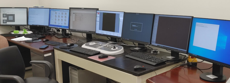
Sifting the data requires graphics cards, and the scientists are competing with people who mine Bitcoin and Ethereum cryptocurrencies, Dr. Olsen said. The price of the cards went through the roof when, after market fluctuations, there was increased mining activity and people were buying loads of graphics cards. “There are supply chain issues, but we’ve gotten what we needed so far,” Dr. Olsen said.
A key addition to UT System cryo-EM capabilities
Much like the space telescopes in Chile, Hawaii and West Texas are indispensable shared resources for astronomers, cryo-EMs are invaluable shared resources for structural biologists. Through its commitment to cryo-EM technology, UT Health San Antonio is entering an arena that will foster interactions within the UT System, where multiple cryo-EMs are in service.
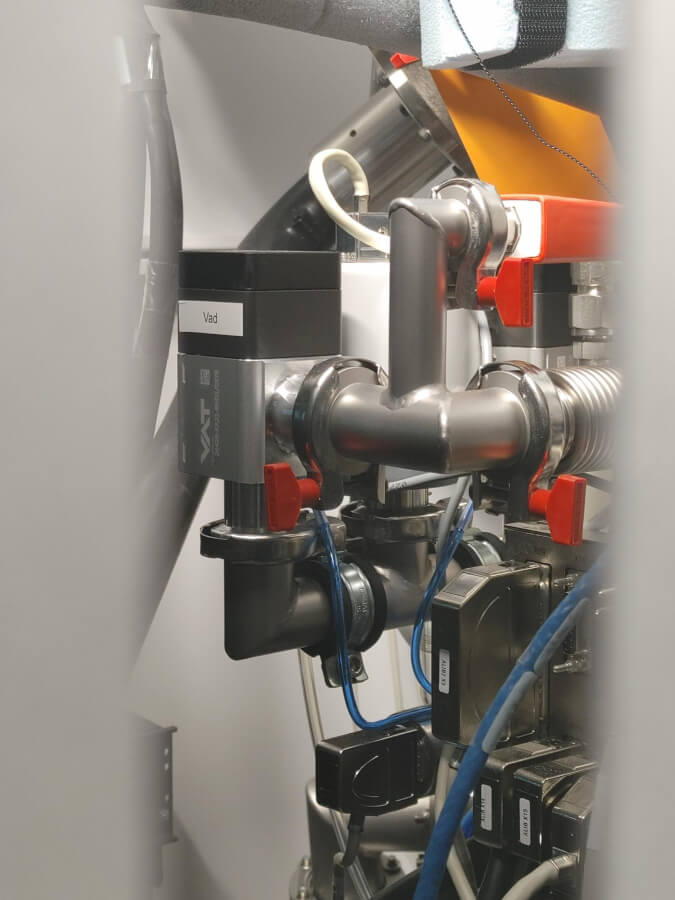
“This microscope will be one of a kind for our area and investigators in South Texas, but it also gives us an opportunity to participate not just by using time at these other facilities, but now we become part of this network. We are all part of the UT System, and we are adding to the capabilities of everyone,” Dr. Olsen said.
The structural biology core at UT Health San Antonio has an agreement in place to pay internal rates to use sister UT institutions’ cores, including their cryo-EMs. UT Southwestern has three, UT Austin has two and UTHealth in Houston has one.
Time is precious, and if there are more microscopes, there is more access and that accelerates everyone’s research.
“These datasets are multi-day,” Dr. Wasmuth said. “You need a lot of data to discover these protein structures. Even though it might seem like there are a lot of cryo-EMs all over Texas, it’s still not enough for investigators here to make the breakthroughs that we need to make.”
Other advantages
To utilize cryo-EM, investigators need protein samples to study. “Our structural biology core facility now has protein production capabilities,” Dr. Olsen said. “If a researcher has a target she is interested in studying, she can come to our scientists to undertake the process of developing samples that are suitable for cryo-EM analysis. If that service is not available, the researcher might never use cryo-EM to ask her question.”
 Many institutions lack this capability, he said. “Basically, we are opening doors for labs that know the important biologic questions but otherwise would not be able to do the structural biology. So, we are providing them avenues to make discoveries,” Dr. Olsen said.
Cryo-EM capability could aid the acquisition of other instruments that will raise up science in San Antonio. With a $350 million annual research portfolio, UT Health San Antonio is the city and region’s largest research university by a significant margin.
The National Institutes of Health’s Office of Research Infrastructure Programs, through its S-10 Instrumentation Grant Programs, supports acquisition of the latest instruments on the market to enhance NIH-funded research nationwide. “Having cryo-EM capabilities on campus should help push our S-10 applications over the top to get funded,” Dr. Olsen said. “It should also help researchers applying for individual NIH research grants, which are called R-01s.”
In capable hands: Dr. Jia, a facility director with rare training in cryo-EM
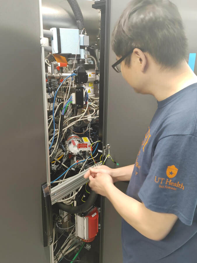
In the new cryo-EM facility, Lijia Jia, PhD, manages a NASA-quality control room with multiple computers, monitors and the cryo-EM system, which includes an electron gun that aims the beams at flash-frozen protein samples.
Dr. Jia is in high demand for his rare skill set in cryo-EM. Specially trained in New York on all aspects of the system, he possesses the crucial ability to precisely align the microscope for target acquisition. He can troubleshoot any issues and knows how to process data and prepare samples. “We are very, very fortunate to have him,” Dr. Wasmuth said. “He is a five-million-dollar man.”
Dr. Jia said he joined UT Health San Antonio because he was impressed by the quality of the science occurring in the institution. The opportunity to establish the new cryo-EM facility was too tempting to pass up, and he spoke about the complementary structural biology techniques that are a part of UT Health San Antonio’s tradition.
A new day in San Antonio
Cryo-EM as a scientific tool has boomed in the last decade due to advances in technology. Better microscopes, better optics and better computer programming have allowed new discoveries. UT Health San Antonio’s cryo-EM system, a Glacios outfitted with a cutting-edge detector (Falcon 4) and energy filter (Selectris), will help investigators of the Mays Cancer Center study tumors. It will help investigators of the Glenn Biggs Institute for Alzheimer’s and Neurodegenerative Diseases to study dementia. It will help faculty of the Sam and Ann Barshop Institute for Longevity and Aging Disorders to study age-related diseases. And, among many other disciplines and applications at UT Health, it will also help researchers of the Greehey Children’s Cancer Research Institute to study childhood cancers.
 “We have to remain competitive, and this is a question that a lot of academic medical centers are trying to decide right now: Where will we fit within the research environment?” Dr. Potter said. “This acquisition reflects our commitment to making San Antonio a biomedical hub for the United States and the world. The types of visualizations and the questions that this technology advances are investments in the long-term improvement of human health.”
Read San Antonio Express-News coverage of this story.
The University of Texas Health Science Center at San Antonio (UT Health San Antonio) is a primary driver for San Antonio’s $42.4 billion health care and biosciences sector, the city’s largest economic generator. As the largest research university in South Texas, with an annual research portfolio of approximately $350 million, UT Health San Antonio drives substantial economic impact through its five professional schools, a diverse workforce of 7,200, an annual operating budget of more than $1 billion and a clinical practice that provides more than 2 million patient visits each year. Furthermore, UT Health San Antonio plans to add more than 1,500 higher-wage jobs over the next five years to serve San Antonio, Bexar County and South Texas. To learn about the many ways “We make lives better®,” visit www.uthscsa.edu.
Stay connected with The University of Texas Health Science Center at San Antonio on Facebook, Twitter, LinkedIn, Instagram and YouTube.
