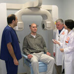
San Antonio (April 27, 2004) – Dental health care has become more sophisticated, more efficient and less expensive at The University of Texas Health Science Center at San Antonio. Thanks to the efforts of Doss McDavid, Ph.D., a faculty member in the department of dental diagnostic science, faculty members at the university are thrilled to be on the receiving end of a new 3-D Accuitomo Cone Beam Micro CT imaging system, worth approximately $200,000. The imaging system, distributed by J. Morita USA, based in Irvine, Calif., arrived Feb. 16. The Health Science Center is one of only three universities in the nation and the only public university to have this equipment. The other two schools are The University of the Pacific in San Francisco and The University of Southern California in Los Angeles.
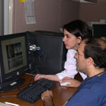
The machine is a dream come true for faculty and students in the Dental School, especially since in February the school was accredited by the American Dental Association’s Commission on Dental Accreditation to train oral and maxillofacial radiology specialists. Dental School faculty and students can view never-before-seen, high-resolution CT images of the jaws to detect implants, abscesses, impacted teeth and other jaw disorders in patients. Using specialized computer software, dentists can view images of jaw structures in three layers simultaneously.
The images can be converted into three-dimensional, life-sized images of the mouth. With the 3-D images, dental radiologists can create life-sized wax models in order to see tumors in the jaws, for example, and measure with exact precision how much tissue from a person’s mouth would need to be removed and/or replaced during surgery.
“This machine allows us to see what’s going on in a patient’s mouth with nearly 10 times the resolution that regular CT machines deliver,” said Robert Langlais, D.D.S., professor and director of the graduate oral and maxillofacial radiology program in the department of dental diagnostic science at the Health Science Center.
“We can see more detail than ever before,” he said. What is even more amazing, Dr. Langlais said, is that the machine can achieve precise images in record time with less radiation exposure to the patient.
“Regular CT machines achieve images by using a beam of radiation that rotates around a patient multiple times, depending upon the part(s) of the jaw or body being scanned,” Dr. Langlais said. “Our new Accuitomo imaging system can achieve a complete scan of the desired part of an individual’s jaw, for example, with one quick beam in about 17 seconds at up to 100 times less radiation dose to the patient,” he said.
Not only does this mean less time, less radiation and more precise images, Dr. Langlais said, it also means less cost to the patient. Regular CT imaging of the jaw costs anywhere from $600 to $800. An Accuitomo imaging session at the Health Science Center costs from $200 to $300.
Dr. Langlais said the technology is so novel to San Antonio and South Texas that the machine at the Health Science Center is becoming very popular. “We have visitors from other departments at the Dental School and from the community almost on a daily basis who want to see this latest technology,” he said. Dr. Langlais said he and other faculty have initiated discussion with faculty members in the School of Medicine at the Health Science Center to apply the technology to others areas of health care.
“Soon a wide variety of applications in all areas of dentistry will be realized, with additional applications in medicine as well,” Dr. Langlais said.