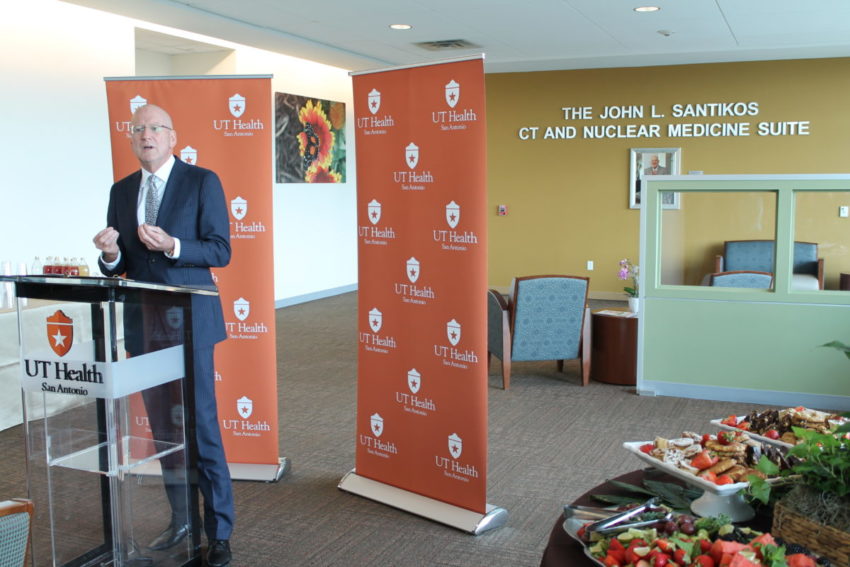
William L. Henrich, M.D., MACP, president of UT Health San Antonio, recognized the John L. Santikos Charitable Foundation, a fund of the San Antonio Area Foundation, on Oct. 2 for its $2.3 million gift to build the John L. Santikos CT and Nuclear Medicine Suite at the university.
The state-of-the-art imaging suite is on the third floor of the Medical Arts & Research Center (MARC) on Floyd Curl Drive. The MARC is a primary location of the UT Health Physicians practice, and the Santikos Suite serves the practice’s patients, including those referred from the community and those of the UT Health Cancer Center.
“This is what success looks like,” President Henrich said in discussing the importance of the Santikos Suite to multiple initiatives such as the new Glenn Biggs Institute for Alzheimer’s & Neurodegenerative Diseases.
Fighting against Alzheimer’s
The community banded together to raise more than $50 million for the Biggs Institute in three years, President Henrich said. A widely published star researcher, Sudha Seshadri, M.D., of Boston University, has been recruited to be the institute’s founding director, effective Dec. 1.
Because of the Santikos gift, the suite named for Mr. Santikos is already serving hundreds of patients who face numerous conditions, such as dementias including Alzheimer’s disease. Treatment to slow the disease is beginning sooner for some patients as a result.
President Henrich thanked Mrs. Ann Biggs and her son, Brian, who attended the event; board members of the San Antonio Area Foundation; and Ed and Nancy Kelley, longtime UT Health San Antonio supporters. Mr. Kelley is immediate past chair of the UT Health Development Board and was involved in establishing the Biggs Institute.
Glenn Biggs, founding chair of the Development Board, was diagnosed with Alzheimer’s in his last years. His need for a comprehensive center to treat and study Alzheimer’s in San Antonio inspired the founding of the institute that bears his name.
Santikos tickets raise funds
Glenn Biggs was a friend of Mr. Kelley’s and would appreciate the development of both the Biggs Institute and the Santikos Suite, Mr. Kelley said. Another friend, John Santikos, specified areas he wanted to support, and a dividend from concessions at Santikos theaters provides philanthropic funding to worthy causes, Mr. Kelley said.
He said a third friend, Sam Barshop, would also appreciate what is happening, especially given the future interactions of the Sam and Ann Barshop Institute for Longevity & Aging Studies with the Biggs Institute.
Aging and risk
Aging is a central risk factor for neurodegenerative diseases. Alzheimer’s disease accounts for 60 percent to 80 percent of dementia cases, and the incidence increases with age. One in 10 people over age 65 will be diagnosed with Alzheimer’s, and this increases to one in three people over age 85.
Alzheimer’s is the sixth-leading cause of death in the U.S. and takes a considerable financial toll on the country. Overall care expenses for Alzheimer’s and other dementias surpass $200 billion annually in the U.S.
Sophisticated instrumentation
The John L. Santikos CT and Nuclear Medicine Suite provides high-resolution:
— computed tomography (CT) and magnetic resonance imaging (MRI), both of which image anatomical structure;
— functional magnetic resonance imaging (fMRI), which enables radiologists to assess blood flow and other properties;
— single photon emission computed tomography (SPECT-CT) and positron emission tomography (PET-CT). In PET-CT, a radiotracer agent is injected into a patient and tracked as it is metabolized in the body.
Clinical trials
In September, a committee of the National Institute on Aging and the Alzheimer’s Association recommended that researchers use biological markers of Alzheimer’s disease, such as brain imaging, when seeking participants for clinical research studies of new therapies.
Researchers in the Joe R. & Teresa Lozano Long School of Medicine at UT Health soon will begin five studies of patients imaged in the Santikos Suite, said Pamela Otto, M.D., professor and chair of radiology who occupies the Stewart R. Reuter Chair in Radiology.
These studies will use the PET-CT camera and newly approved PET brain imaging agents to diagnose and monitor amyloid plaque in Alzheimer’s disease, Dr. Otto said.
Prevention not yet a reality
“It would be best to prevent the disease so that people never develop it,” she said in an interview prior to the Santikos Suite ceremony. “We don’t have that yet, but the next best alternative is early detection and treatment, because right now all we can do is slow progression.”
Because there are multiple types of dementia, a hurdle to doing such studies is making sure the patients enrolled actually have the particular dementia to be studied, Dr. Otto said.
Different disease, different set of images
“The different neurodegenerative processes hit different areas of the brain,” she said. “Alzheimer’s looks different than Lewy body dementia. Frontotemporal dementia looks different than vascular dementia. Depending on the imaging characteristics, we can pinpoint which one.”
For example, by using a radiotracer agent in the PET-CT camera, the radiologists can see if no glucose is being metabolized in the temporoparietal region – a deficit that is very common in Alzheimer’s disease, Dr. Otto said.
Credentials
The Santikos Suite is fully credentialed to conduct studies in Alzheimer’s, cancer and other diseases. On the topic of cancer, Dr. Otto said the Santikos Suite can do every imaging study that any other major cancer center in America can do.
Rajeev Suri, M.D., professor of radiology, said CT, in addition to its role in Alzheimer’s, helps in the staging of cancer treatment, evaluating metastasis (cancer spread) and tracking of treatment effect.
Based on structural imaging alone, oncologists and other members of a multidisciplinary treatment team may only be able to see a tumor shrink over time, possibly as long as a year, Dr. Suri said.
Structure and function in cancer imaging
“With PET-CT, you can see actively dividing cells using up the glucose,” he said. “You can actually learn that the activity of the cancer is gone, and be confident that this will be followed, in a matter of time, by tumor shrinkage.”
Umber Salman, M.D., associate professor of radiology and director of nuclear medicine, said radiologists do a baseline set of imaging studies for patients and follow up between treatments to see if a therapy is working. Patients are having follow-up imaging studies sooner nowadays—in half the time in some cases. For many cancers, the follow-up imaging generally occurs in three to six months. PET-CT and radiotracers are accelerating that, which means patients who aren’t responding to therapy can be switched to another therapy faster.
“The interval between the baseline and follow-up studies is different for every tumor, because each responds differently to treatment depending on its type and its level of aggressiveness,” Dr. Salman said.
The University of Texas Health Science Center at San Antonio, with missions of teaching, research and healing, is one of the country’s leading health sciences universities and is now called UT Health San Antonio™. UT Health’s schools of medicine, nursing, dentistry, health professions and graduate biomedical sciences have produced more than 33,000 alumni who are advancing their fields throughout the world. With seven campuses in San Antonio and Laredo, UT Health San Antonio has a FY 2018 revenue operating budget of $838.4 million and is the primary driver of its community’s $37 billion biomedical and health care industry. For more information on the many ways “We make lives better®,” visit www.uthscsa.edu.