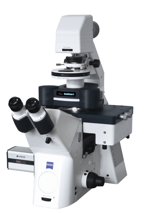
The University of Texas Health Science Center at San Antonio (also called UT Health San Antonio) received a grant from the National Institutes of Health in excess of $590,000 that will be used to purchase a NanoWizard® 5 Atomic Force Microscope (NW5 AFM), the first of its class to be installed in San Antonio.
The custom-built microscope, the newest state-of-the-art instrument, is slated to be installed later this year. It will join a Catalyst AFM microscope that, over the last decade, has supported breakthrough research of biophysical properties of single cells, including cells isolated from cancer patients.

“When we applied for this grant last year, there was no instrument like it installed in the world,” said Pawel Osmulski, PhD, associate professor of research in the Department of Molecular Medicine in the Joe R. and Teresa Lozano Long School of Medicine. Osmulski is the grant’s principal investigator.
“The capabilities of this new AFM will allow even deeper development of hyphenated approaches to study fundamental questions in biomedicine and biomaterials,” Osmulski said. “It is our pleasure to report a significant expansion of cutting-edge research capabilities of the AFM Laboratory, which is part of the institution’s Bioanalytics and Single Cell Core (BASiC), directed by Nameer Kirma, PhD, associate professor in the Department of Molecular Medicine.”
The biophysical arm of services of the BASiC Core is provided by the AFM Laboratory, directed by Osmulski.
The expanded capabilities of the NW5 AFM microscope include the following:
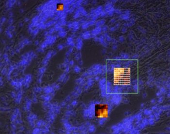
- Providing nanomechanical phenotypes (and fluorescent images) of not only single cells, as the current microscope provides, but also tissue slices and organoids, all at much higher speed and resolution than before.
- Probing direct cell-cell or ligand-cell interactions for a variety of cells, including very large cells inaccessible by the current equipment. This opens a unique opportunity to study cell microenvironments, for example, in cancer or neural tissue.
- Performing sophisticated manipulations on single cells, such as moving cells from one environment to another, non-destructively injecting nano-volumes of material directly into cells or sampling a nano-volume of cell contents for further analysis.
- Performing high-speed and high-resolution molecular-scale imaging of biomacromolecules such as proteins and nucleic acids; such imaging will enable studies of dynamic interactions between biomacromolecules and their ligands.
The new equipment is already attracting an impressive array of multidisciplinary researchers from across the institution and the greater San Antonio research community interested in its future use to elevate the institution’s research enterprise and improve community health in South Texas.