The winners of Briscoe Library’s 3rd Annual Image of Research Photography Competition have been announced.
1st Place
Breeanne Soteros, Graduate School of Biomedical Sciences
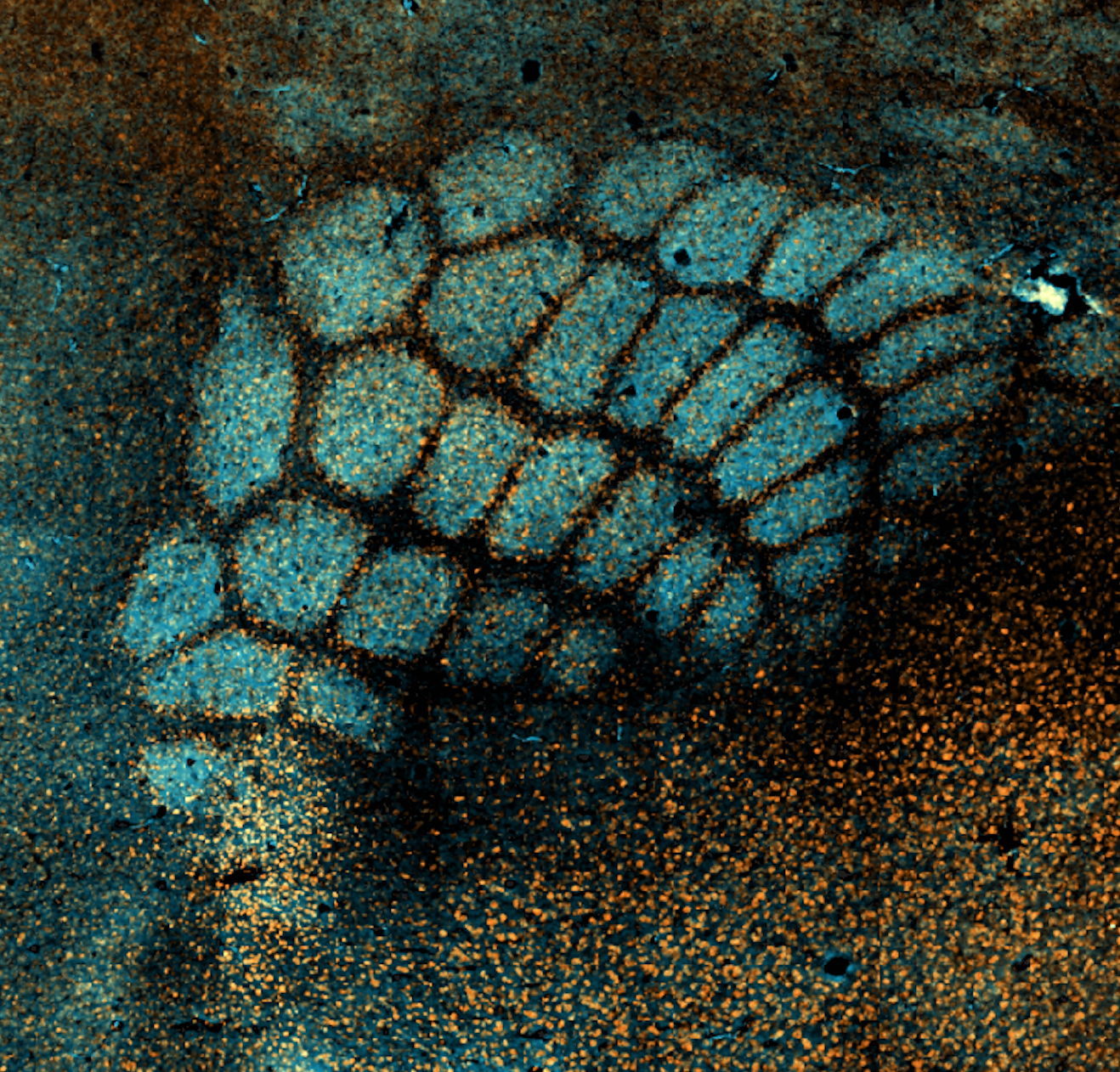
Flattened cortical brain sections stained for glutamate transporter VGlut2 reveals the somatotopic organization of the mouse barrel cortex. Each “barrel” (in blue) corresponds to the major facial whiskers of the mouse, with the topographical organization of the cortex closely resembling the whisker pad itself. Co-staining for the immune protein C1q (in orange) reveals an unexpected pattern – this complement cascade molecule appears to decorate the borders of each barrel. In the human brain, the complement protein C1q is important for shaping the synaptic landscape, as it tags synapses for elimination. Could this immune help define these barrels by eliminating excess synapses at the border? What might this pattern of staining reveal about the ongoing synaptic maintenance of sensory circuits in the brain? Through a combination of genetic and molecular techniques, we hope to tease out the mechanisms of synapse maintenance & elimination that govern the organization of both the mouse and human brain.
2nd Place
Pragya Singh, Graduate School of Biomedical Sciences
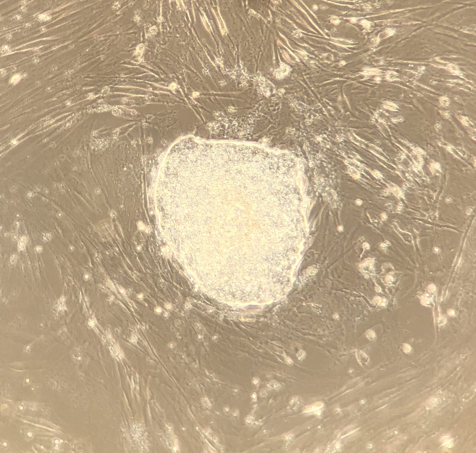
It was early 2018 I had a breathtaking moment when I first saw iPSC clones during my Master’s research here in UTHSA. I was reprogramming fibroblast into stem cell-like cells known as induced Pluripotent Stem Cells or iPSCs. These iPSC clones are so malleable that you can influence any cellular fate you wish to generate. This pristine clone was further induced to generate Retinal Ganglion Cells to understand the molecular mechanism of Leber’s Hereditary Optic Neuropathy, a rare genetic disease that leads to vision loss.
3rd Place
Sonam Khurana, School of Dentistry
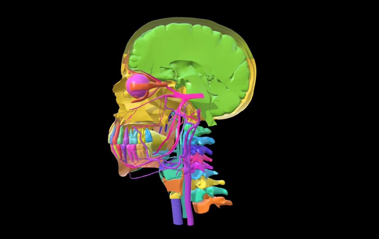
This three-dimensional (3-D) animation you are seeing is depicting nerve and blood supply to the teeth. The model is a complicated piece of art, requiring lots of practice and skills to be produced. 3-D animation has been used in entertainment industry (like in movie theaters!) to create a range of exciting videos, short cartoons and full-length videos. The health care industry also uses 3-D animations create models that everyone can examine. The use of anatomical models is ubiquitous in health education. Realistic looking models allow the user to move away from complex cadaveric dissection, which is not a readily available resource any way. Models are very useful to explain anatomical relationships and function in structures that may be too small to discern adequately in a cadaver. Learning anatomy is challenging, and an adaptation of new methods that are user-friendly is essential. We hope that our research based on creating anatomical models will help the transition from dissection to the use of 3-D animations. It will not only help health professionals to learn anatomy nimbly, but also offer them a tool that is easy to show and explain to their patients. Let the journey begin!
IPE Award
Rafael Veraza (Anesthesiology), Jaclyn Merlo (Immunology and Infection), and Kristina Andrijauskaite (Molecular Medicine)
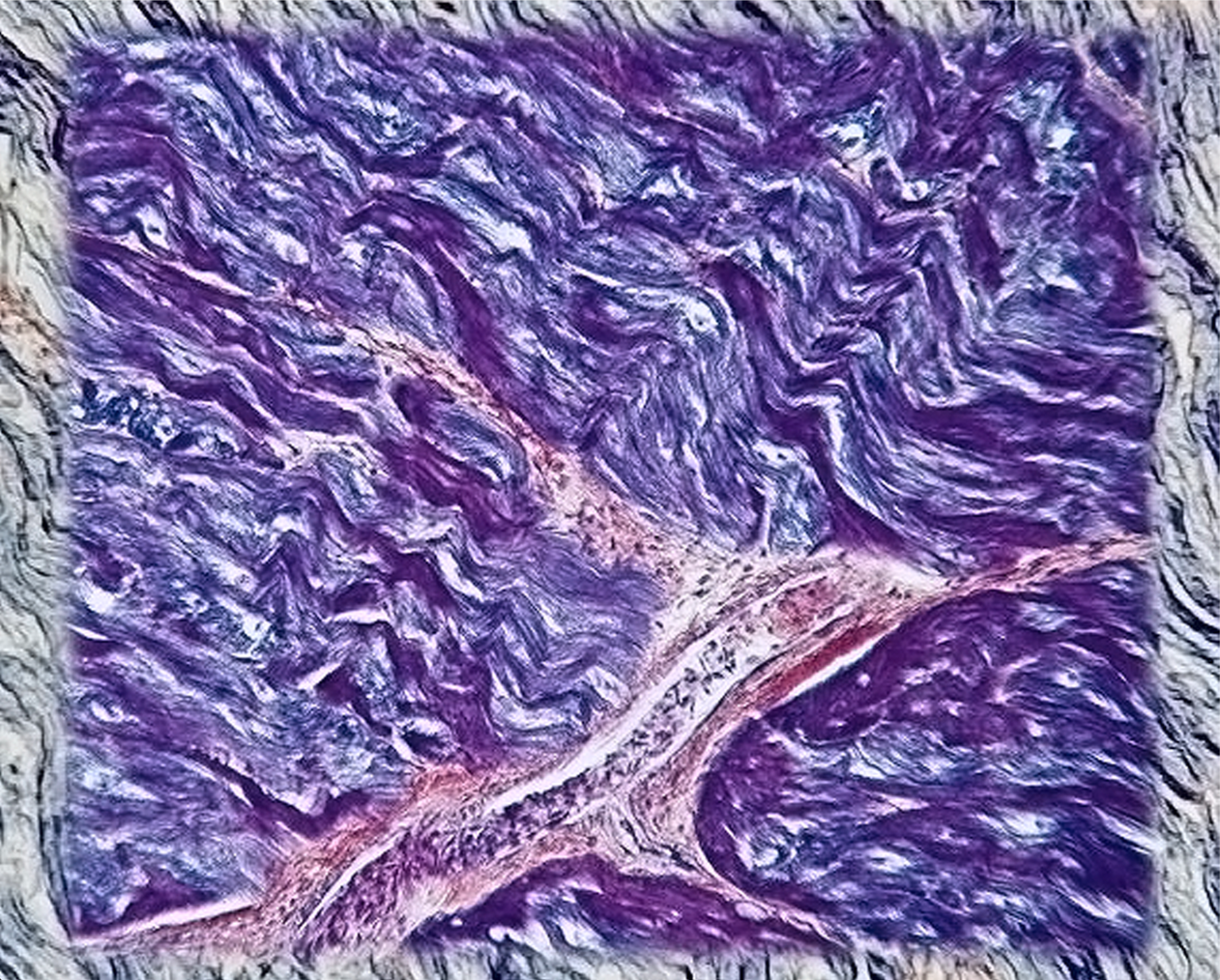
This combined image consists of ischemic cardiac pig muscles overlaid with a blood vessel. The tissue was removed and stained from an ischemic heart placed in cold storage; the traditional method of heart preservation for transplantation. Our aim is to extend organ tissue preservation beyond the time constraint of 4 hours via novel biomedical devices. This will increase the viability of organs for transplantation beyond the current standard of care. The BLUE heart represents the lack of time in the field of tissue preservation. BLUE encompasses our limitations on delivering a warm RED heart to a transplant recipient. Our multidisciplinary group preserves tissues of the heart, colon, and limbs for periods of 24 to 48 hours outside of the body and studies hypoxia makers to further improve transplant outcomes.
Faculty/Staff Award
Sang Hyun (Ryan) Chun, Research Associate, Graduate School of Biomedical Sciences
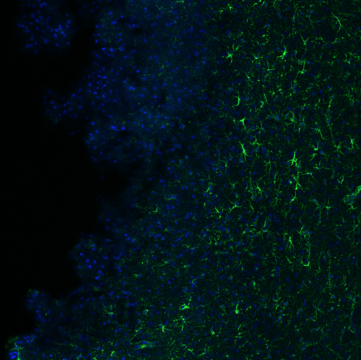
Traumatic Brain Injury (TBI) has been garnering attention as one of the most prevalent neuropathogenesis. Injuries occurring from sports related activities, wars, and domestic violence are the few examples of many TBI related incidents. The underlying mechanism of TBI and its therapeutic intervention have been poorly defined and must be addressed. Astrocytes have been regarded as a key agent in maintaining brain homeostasis. Astrocytes are the most abundant glial cells in our central nervous system. As the name implies, astrocytes are known for having a star shaped characteristic in the brain. In addition to sustaining brain homeostasis, astrocytes have shown key neuroprotective role after TBI. After injury, the astrocytes undergo what is known as astrogliosis in which morphological and molecular changes occur. In our study, we use cytoskeletal protein called glial fibrillary acidic protein (GFAP) as a biomarker to look at how the astrocytes function after the injury. The image is displaying a cortical region of the mouse brain after TBI. GFAP labeled astrocytes and nuclei are shown in green and blue, respectively. This image captures how the brain tries to react to the injury by undergoing astrogliosis and the enlarged astrocytic “stars” are being activated as a neuroprotective measure.
All of the submissions that Briscoe Library received were very impressive. The photos were all scored based on visual impact, connection between image and research, and originality by our multidisciplinary panel of judges. While we had initially planned on having an Image of Research Awards Reception this Spring, the reception has been postponed. Please stay tuned for updates.
Click here for more information regarding the competition rules, guidelines and details.

