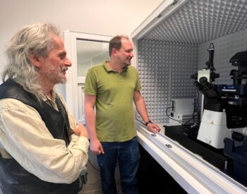
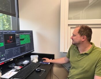
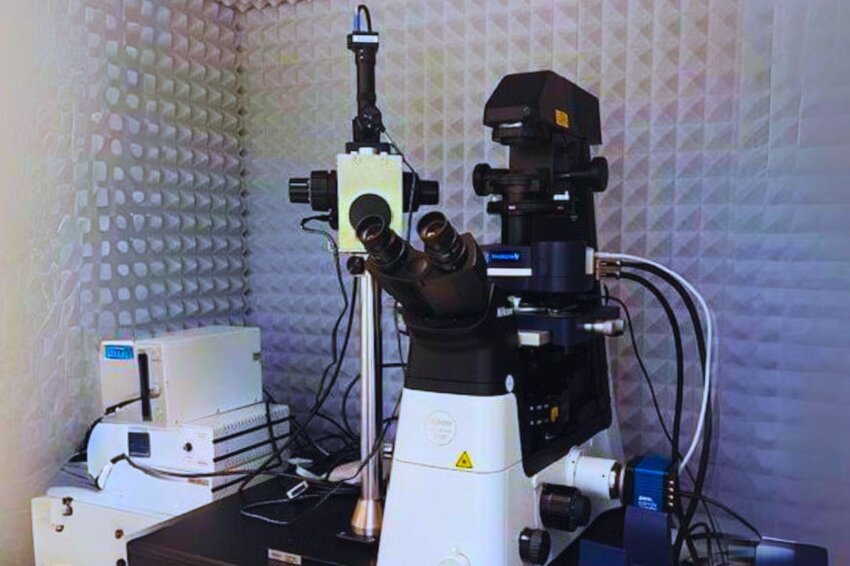
The bioanalytics and single-cell core (BASiC) facility at The University of Texas Health Science Center at San Antonio has a new, first-in-Texas Nanowizard V atomic force microscope.

Pawel Osmulski, PhD, department of molecular medicine, took the lead in applying for a National Institutes of Health grant (S10 funding) for the specialized equipment. Nameer Kirma, PhD, director of BASiC, and Maria Gaczynska, PhD, assisted in the effort.
UT Health Science Center San Antonio first acquired an atomic force microscope in 1997 in the department of medicine, then an additional one in 2013 at BASiC, which served the health science center and other scientific communities in Texas and the US. The other atomic force microscope is adequate for most single-cell work, however, the newest cutting-edge instrumentation was necessary to enhance and enable translational research potential.

The NIH’s specialized instrument grant process began in 2022, which involved collecting letters of support from the institution and other researchers, together with descriptions of specific research projects and publications related to the microscope’s capabilities. The team learned the funding was secured in August 2023.
Several university departments supported the effort, including the biochemistry, structural biology and molecular medicine departments, the Greehey Children’s Research Institute, the Barshop Institute for Longevity and Aging Studies, the Mays Cancer Center and the School of Dentistry. UTSA and other UT Systems schools also contributed.
Osmulski and his team were awarded $595,000 in NIH funding for the new microscope. Another $20,000 was crowd-sourced from various health science center researchers to purchase additional equipment to boost the microscope’s abilities even further.

“I am especially looking forward to the equipment’s 3D imaging capabilities and working with tissue slices that we were not able to previously to enable correlative studies that include mechanical properties, protein expression and transcriptomics profiling of the same tissue,” Kirma said.
“One of the nice things about it is we can look at the surface of the cells, whether it’s in vitro or whether it’s tissue sections with the new instrument. So now we’re adding the spatial component to the single-cell analysis,” he added.
The atomic force microscope and related equipment will let the team preserve samples in a stable environment for longer periods of time, allowing them to better study spatial-temporal effects on mechanical properties of a cell.
Kirma’s research focuses on endometrial cancer, and he said the new microscope will allow the study of the interaction of different types of cells such as the adherence of cancer cells onto substrate lining.
For other experiments, researchers can inject a cell with a reagent and introduce it to another cell to detect changes, as well as pull material from one cell and transplant it to another cell.
Osmulski is especially looking forward to the improved speed for molecular studies.
“You can look at how an enzyme is performing its catalysis or how a protein travels on DNA molecules in real-time. With this [new microscope], we can observe this 100 times faster than before,” he said.
Kirma said the microscope is a “gold mine of opportunity” for the scientific community in San Antonio.
“We are able to better explore 3D structures, from tissue to organoids, which was not possible before. That increases the translation aspect of the research,” he said.
The NanoWizard V atomic force microscope has been installed in the university’s South Texas Research Facility on the Greehey Campus.


