Advanced dental imaging is a safe, noninvasive tool that guides health providers and their patients toward sound treatment decisions.
At UT Health San Antonio School of Dentistry’s dental practice, UT Dentistry, Rujuta A. Katkar, BDS, MDS, MS, and her team use the latest imaging technologies to provide accurate diagnoses and treatment planning support for a variety of dental specialties.
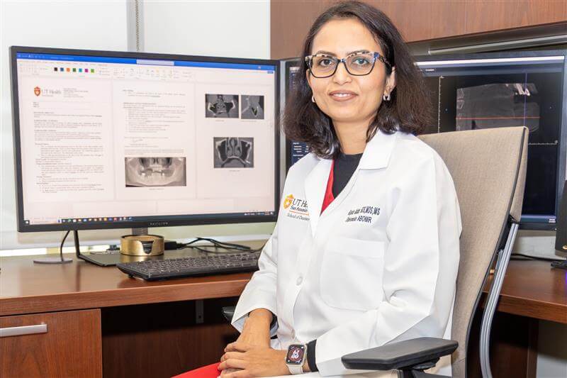
Katkar, an associate professor and board-certified oral and maxillofacial radiologist, is trained in various imaging modalities, including intra- and extra-oral radiography, ultrasound, magnetic resonance imaging and cone beam computed tomography (CBCT) of the bones and tissues of the mouth, jaws and face.
“We spend two to three years in a CODA accredited training program after graduating from a dental school, with a rigorous curriculum focused on imaging techniques, anatomy and pathological conditions of the oral and maxillofacial region,” Katkar said. “We train by interpreting thousands of images, especially CBCT scans, to detect the subtle signs of disease or incidental findings that need to be taken care of or require a referral to a physician.”
Oral and maxillofacial radiology’s role
The importance of oral and maxillofacial radiology (OMR) cannot be overstated, according to Katkar. Her team provides essential support to in-house and referring dental providers by interpreting imaging scans and offering treatment recommendations.
“For example, we assist in planning dental implants or complex oral surgeries by assessing bone quality and proximity to critical structures such as nerves,” Katkar said. “We can also detect pathologies that may go unnoticed with traditional 2D imaging.”
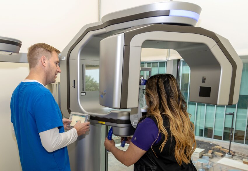
The Oral and Maxillofacial Radiology Clinic is equipped with the latest CBCT machines, which use a cone-shaped X-ray beam that rotates around a patient’s head to produce detailed, three-dimensional images of the teeth, jawbones and surrounding structures.
“This capability ensures more precise diagnoses and treatment plans, ultimately improving patient care,” Katkar said.
While CBCT machines are increasingly common in dental offices, Katkar urges providers to be cautious.
“Proper technique and interpretation of these scans require specialized training,” she said. “If the scans aren’t of good diagnostic quality, a provider may miss important details.”
The difference imaging makes

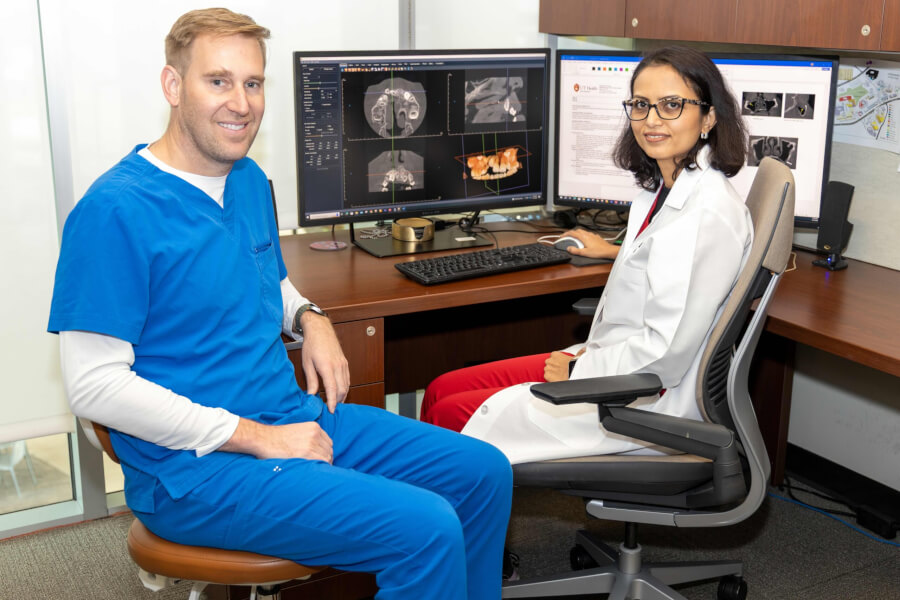
Katkar and her team frequently collaborate with colleagues in general dentistry, endodontics, periodontics, orthodontics and oral surgery to guide clinical decision-making. She said advanced imaging and interpretation has proven crucial in saving patients from unnecessary surgeries or complications.
“For example, we’ve seen several cases where a cyst, tumor or infection was initially missed on a radiograph or CBCT scan acquired by a dentist, only to find out later that it was right there on all the prior images, but went unnoticed,” she said.
Another case the OMR team consulted on involved a patient who had persistent temporomandibular joint pain. The source of the pain was eventually traced to a tumor, which was only visible through advanced imaging.
“These are the cases where advanced imaging truly makes a difference. Without it, we might miss significant pathologies that could affect a patient’s health long-term,” Katkar said.
While some providers may prefer not to send every scan to a radiologist for interpretation, Katkar urges them to contact the clinic if they are ever uncertain.
“Oral and maxillofacial radiologists are often called on to serve as legal experts — that’s how much weight a scan interpretation can carry,” she said. “Providers are responsible for the entire volume of a scan they acquire and will be held to the same standards as a specialist in the court of law, so it’s important for them to consult us if something looks suspicious — we are here to help.”
Katkar and her team also offer dental providers guidance on obtaining the best quality images for patients’ specific diagnostic needs, taking into consideration radiation safety and quality assurance for compliance with state regulations.
AI and dental imaging
The field of oral and maxillofacial radiology is constantly evolving with advancements in imaging technologies and techniques, including the integration of artificial intelligence (AI) into diagnostic tools and 3D printing for surgical guides.
“AI is in the testing phase, and while it can be a valuable second opinion, we need to ensure its accuracy before relying on it for diagnosis,” Katkar said.
She added that the OMR team at UT Dentistry is committed to contributing to this research for the field’s growth to verify that these tools are developed safely and effectively.
“Our goal is to provide top-tier diagnostic services, so we must contribute to the continuous advancement of our field,” Katkar said. “We are excited about what the future holds and the opportunities it brings.”
To learn more about UT Dentistry’s Oral and Maxillofacial Radiology Clinic, visit their website or call 210-450-3220.